| 11444-000 | light tone | Quote |
|---|---|---|
| 11444-000-M | medium tone | Quote |
| 11444-000-D | dark tone | Quote |
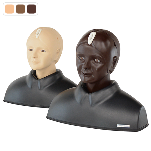
Innovative Eye Examination Simulator.
- OSCE
- Ophthalmoscopy
- Nurse Practitioner
- General Practitioner
- Family Medicine
MW61/MW61A
| 11444-000 | light tone | Quote |
|---|---|---|
| 11444-000-M | medium tone | Quote |
| 11444-000-D | dark tone | Quote |
Innovative Eye Examination Simulator.
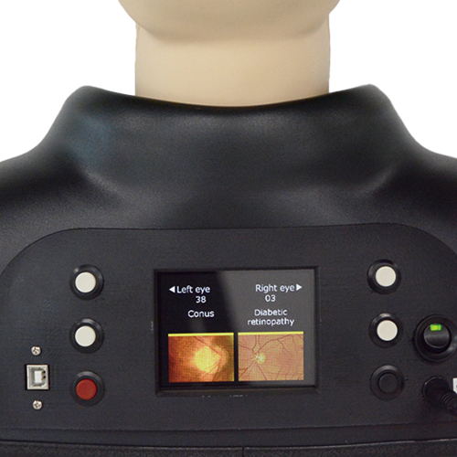
| Features | Our standard Eye Examination Simulators are utilized by many simulation centers. This digital version of the Eye Examination Simulator is an upgrade to showcase various innovative features. 1. 40 cases of fundus images (including 10 common diseases) are pre-installed for eye examination training and viewable on 160dpi monitor 2. Pre-installed fundus images are from actual cases using real clinical images in order to properly reproduce appropriate representative views of the eye unit 3. Custom fundus images can be programmed into the simulator via USB 4. Lens-equipped eyeball units that simulate the visual axis of the human eye can be examined using all types of direct opthalmoscopes 5. Soft and supple material, allowing for realistic raising of the eyelid 6. Changing the degree of dilation and contraction of the pupil in 3 steps (2, 3.5,5mm) offers different degrees of challenges. *MW61A 11444-999 is 2 steps; 3.5, 8mm) |
|---|---|
| Training skills / Applications | Fundus examination with real ophthalmoscope |
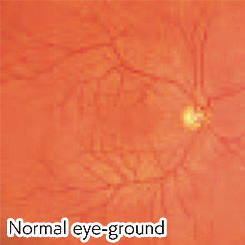
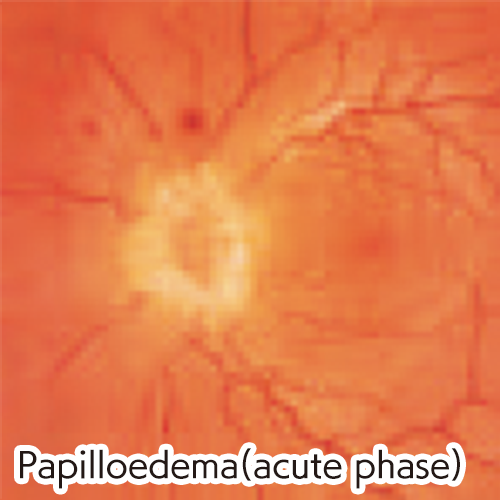
| Case / Pathology | Normal eye-ground / Hypertensive retinopathy: arteriolar vasoconstriction grade 3, arteriolosclerosis grade 1, hemorrhages and cotton wool spots, simple vein concealment / Simple/background diabetic retinopathy: microaneurysm, hemorrhages and hard exudates / Papilloedema (chronic phase) / Papilloedema (acute phase) / Glaucomatous optic atrophy: glaucomatous optic disc cupping and nerve fiber defect / Retinal vein occlusion (acute phase): flame-shaped hemorrhage and cotton wool spots / Retinal vein occlusion (after retinal laser photocoagulation) / Toxoplasmosis: retinochoroiditis / Age-related macular degeneration: macular exudates and subretinal hemorrhage Custom fundus images can be installed on simulator via USB |
| Set includes | 1 manikin head and shoulder / 1 pupil switch- 3 steps (2, 3.5, 5mm diameter) - 2 steps (3.5, 8mm dia.) *MW61A / 1 power cable / 1 USB cable / 1 USB flash drive (with case installer) / 1 instruction manual |
| Size (approx.) | W42 x D21.5 x H38 cm / W16.5 x D8.5 x H15 in |
| Packing size (approx.) | W46 x D30 x H54 cm / W18.1 x D11.8 x H21.2 in |
| Weight (approx.) | 2.5kg / 5.5lbs |
| Packing weight (approx.) | 4.35kg / 9.6lbs |
| Power requirements (approx.) | 2.5W |
| Materials | Soft resin / Hard resin / Latex free |
| Related products |
Eye Examination Simulator
|
| Different sizes of pupil | It is possible to change the degree of dilation and contraction of the pupil. MW61 3 steps: 2, 3.5, 5mm 11444-000 11444-000-M 11444-000-D MW61A 2 steps: 3.5, 8mm 11444-999 11444-999-M 11444-999-D |
| CE certified/CE Certification? | Yes |
| Update | May 27, 2025 |