Digital EAR Examination Simulator II
Product Supervision
Japan Society for Medical Education
with the cooperation of Michihiko Sone Professor - Nagoya university Graduate school of Medicine
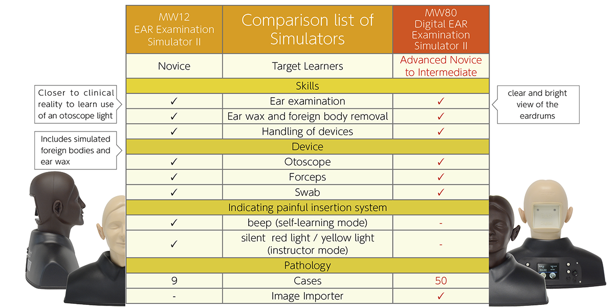
Comparision
50 Pre-set Cases

-- Cases No.10–50: All-New Content --
Normal
- 01~04. Normal tympanic membrane (4 patterns)
- 05~06. Normal tympanic membrane (pediatric) (2 patterns)
Outer Ear
- 07~08. Cancer of external auditory canal-adenoid cystic carcinoma (2 patterns)
- 09. Cerumen block
- 10. Earwax
- 11. External auditory canal cholesteatoma
- 12. External auditory canal exostosis
- 13. Medial meatal fibrosis
- 14. Traumatic tympanic membrane perforation-post myringoplasty
- 15. Seborrheic keratosis of the external canal
- 16. Acute otitis media
Middle Ear
- 17. Acute suppurative otitis media(AOM)
- 18. Aerotitis media after myringotomy
- 19~22. Cholesteatoma (4 patterns)
- 23. Cholesterol granuloma of the middle ear
- 24. Chronic otitis media
- 25~28. Chronic otitis media with perforation (4 patterns)
- 29. Eosinophilic otitis media
- 30. Glomus tympanicum tumor
- 31. Large tympanic perforation
- 32. Lateral tympanoplasty
- 33. Mucoid otitis media(MOM)
- 34. Otitis media with effusion
- 35. Retracted tympanic membrane
- 36. Serous otitis media(SOM)
- 37. Traumatic tympanic membrane perforation (2 patterns)
- 38. Tuberculous otitis media
- 39. Tympanic membrane atelectasis
- 40. Tympanosclerosis
Inner Ear
- 41. Cochlear otosclerosis
Congenital Case
- 42. Congenital ossicular anomaly-malleus bar
Pediatric
- 43. Adhesive otitis media (pediatric)
- 44. Chronic otitis media with perforation (pediatric)
- 45~46. Keratin cyst of the tympanic membrane (pediatric) (2 patterns)
- 47 Otitis media with effusion (pediatric)
- 48. Tympanic membrane atelectasis (pediatric)
After Surgery
- 49. Postoperative ear (cholesteatoma)
- 50. Tympanostomy tube (pediatric)
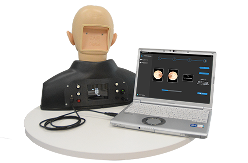
Image Importer
Install Your Original Images
In addition to the 50 pre-installed images, you can import up to 50 of your own images into the simulator via USB, using the case installer included in the set.
User-Friendly Design
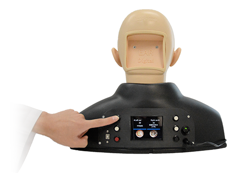
New design that is convenient for both instructors and learners.
- The case images can be switched with a single click of a button.
- The shown case can be monitored on the rear screen.
- Different cases can be displayed on the left and right ear.
Other Features (Shared with MW12)
- Anatomically correct soft ear model
- 2 sizes of ear canals (normal and stenosis)
- Ear wax and foreign body removal
- Training with a real otoscope