| 13150-000 | 936,430 (税別851,300)円 |
|---|
- 解剖模型
- 心臓
- 肺
- 3Dプリント
EZ-151
| 13150-000 | 936,430 (税別851,300)円 |
|---|
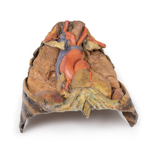
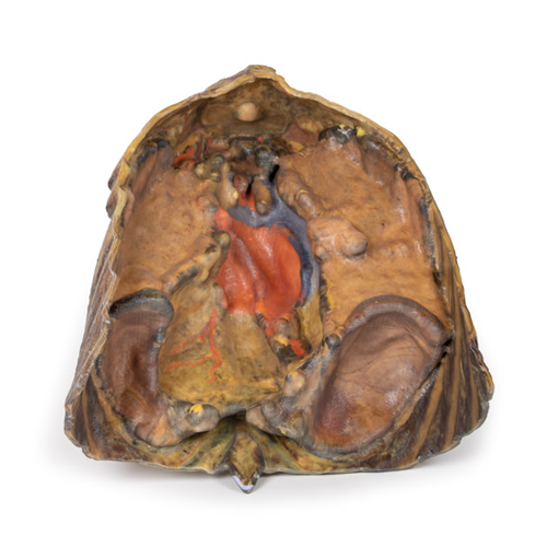
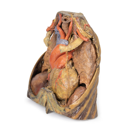
| 特長 | 本製品の説明は英語のみです。 Product information "Thorax with heart and vessels" The superior thoracic aperture contains structures emerging from the thorax and entering the head and neck and upper limb. In this specimen, both clavicles, key venous structures and other musculature have been removed. Despite this, other important components of anatomy can be observed. Key structures include the Trachea seen most superiorly with a thick ring of cartilage, rib one has been exposed prior to meeting its costal cartilage, travelling in a lateral to medial direction and the anterior scalene muscle inserting into Rib one superiorly. In regards to the blood supply, The right subclavian can be seen superior to rib one, giving off the thyrocervical trunk to supply the neck, the left subclavian can also be seen superior to rib one, giving off the suprascapular artery and both the left and right common carotid can be seen superiorly with the left also containing a clear left vagus nerve. Travelling inferiorly from the cranium, the vagus nerve follows the common carotid arteries in the carotid sheath which can be seen in the left common carotid artery. The left phrenic nerve remains unclear until it emerges in the mediastinum. Components of the left brachial plexus can be seen, from roots to trunks. Numerous smaller branches move off, including the Dorsal Scapular nerve. In the mediastinum, key structures can be identified in the mediastinum of the thorax. However, other pieces of anatomy including the inferior vena cava, right vagus nerve and some coronary vessels cannot be observed in this specimen. The Right phrenic nerve can be traced posteriorly to the heart, this has been shifted from its normal anterior to the heart position during dissection. Also seen is the Left phrenic nerve is still contained in its connective tissue and remains located anteriorly to the heart, until reaching the diaphragm for innervation. The left vagus nerve can be seen posterior to the heart and is easier to identify superiorly following the left common carotid artery in the carotid sheath. Note the left recurrent laryngeal nerve moving under the aorta. The Arch of aorta giving branches of brachiocephalic and left common carotid superior to the heart. The left subclavian artery can be seen just posterior to the left common carotid. With the pulmonary trunk exiting immediately superior to the heart. The Left anterior descending carotid artery can be observed cascading anteriorly on the heart with the superior vena cava can been seen to the right of the aorta and posterior. The inferior thorax shows Ribs 8 through 12 on the specimen, the musculature in-between these ribs can also be seen. Notably, the direction of external intercostal muscle inferomedially can be seen as it progresses into a layer of fascia. As observed in the specimen, the right hemidiaphragm is located more superiorly than the left hemidiaphragm, and this is due to the presence of the liver on the right side of the abdominal cavity. |
|---|---|
| 仕様 | エルラージマー社製 |
| 消耗品購入 | |
| 備考 | メーカー品番:MP1122 Erler-Zimmer GmbH & Co.KG の模型製品は、日本国内において株式会社京都科学の独占販売製品です。 |
| 医療機器クラス分類 | なし |
| 特定保守管理医療機器 | 該当なし |
| JANコード | なし |
| 更新年月日 | 2020年06月14日 |
