| 13156-000 | 2,492,600 (税別2,266,000)円 |
|---|
- 解剖模型
- 腸
- 胴体
- 3Dプリント
EZ-157
| 13156-000 | 2,492,600 (税別2,266,000)円 |
|---|
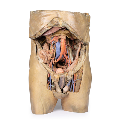
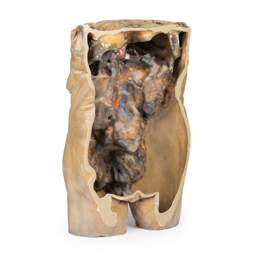
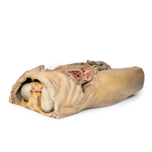
| 特長 | 横隔膜から大腿部までの詳細な構造を再現。鼠径ヘルニアを左右同時に発症した希少な献体を解剖。 Product information "Abdomen with bilateral Hernias" This 3D model represents one of the largest and most complex in the series, consisting of a partial torso from the diaphragm to the proximal thigh with a complete abdominal cavity preserving varying levels of dissection. This 3D model also records the rare, simultaneous occurrence of indirect and direct inguinal hernias allowing for a consideration of the anatomical underpinnings for both conditions. Given the scale of the dissection this 3D model description is divided into discrete parts based on views and regions. The diaphragm On the superior aspect of the model the diaphragm is preserved, and while slightly distorted due to removal of the thoracic ribs through dissection, both domes and costodiaphragmatic recesses can be appreciated. The fibrous pericardium is present on the superior surface of the central tendon, with the terminal part of the inferior vena cava visible in the caval foramen. Just lateral to caval foramen is the oesophagus within the oesophageal hiatus, and then the descending thoracic aorta approaching the aortic hiatus just ventral to the thoracic vertebrae. The epigastric and hypochondriac regions Within the abdomen, the anterior abdominal wall, greater omentum, and much of the gastrointestinal tract has been removed alongside the parietal peritoneum over the posterior abdominal wall to expose retroperitoneal organs and structures. In the superior abdomen, the terminal portion of the oesophagus has been retained and can be seen entering the cavity just lateral to the left lobe of the liver. The removal of the stomach has exposed the extent of the pancreas from the head (positioned within the arc of the duodenum) to the tail extending to the capsule of the spleen preserved in the left hypochondrium. Superior to the pancreas, the splenic artery and common hepatic arteries can just be observed spanning across the narrow space between the pancreas, diaphragm and liver. The splenic follows its archetypical ‘tortuous route’ towards the spleen, and strongly divides prior to reaching the hilum (and adjacent to the splenic vein). The common hepatic can be seen dividing into the gastroduodenal (visible again as a cut vessel just inferior to the duodenum) and giving off the right gastric artery; these vessels lie superficial relative to the hepatic portal vein. The superior mesenteric artery and vein can be seen passing anteriorly near the head of the pancreas and horizontal part of the duodenum, and the retained ileocolic artery can be traced to the caecum of the large intestine in the lower right quadrant of the abdomen. The inferior mesenteric vein can be, in part, appreciated arising from the retained superior rectal vein ascending from the undissected true pelvis and spanning across the superficial aspect of the descending thoracic aorta. Inferior to the liver the gallbladder can be viewed just between the right and left anatomical lobes. On the left, the passage of the renal artery and vein can be seen just deep to the pancreas, and the ureters can be observed descending from the partially exposed kidney across the superficial surface of the exposed psoas major and minor muscles. The umbilical and lumbar regions Most of the organs occupying the umbilical and lumbar regions of the abdomen have been removed in order to expose structures in the posterior abdominal wall. In the midline, the descending abdominal aorta and inferior vena cava dominate the region, with the testicular arteries and veins isolated and traceable towards the inguinal regions. Two right lumbar arteries are visible arising from the aorta, and despite removal of the mesenteries and most of the colon the inferior mesenteric artery can be seen giving rise to the left colic, sigmoid and superior rectal arteries. On the right side of the specimen inferior to the kidney, the subcostal, iliohypogastric and ilioingual nerves are exposed alongside the circumflex iliac artery. |
|---|---|
| 仕様 | エルラージマー社製 |
| 消耗品購入 | |
| 備考 | メーカー品番:MP1130 Erler-Zimmer GmbH & Co.KG の模型製品は、日本国内において株式会社京都科学の独占販売製品です。 |
| 医療機器クラス分類 | なし |
| 特定保守管理医療機器 | 該当なし |
| JANコード | なし |
| 更新年月日 | 2020年06月11日 |