| 13165-000 | 1,286,120 (税別1,169,200)円 |
|---|
- 解剖模型
- 腹部
- 下半身
- 3Dプリント
EZ-166
| 13165-000 | 1,286,120 (税別1,169,200)円 |
|---|
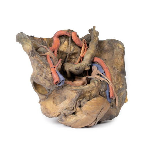
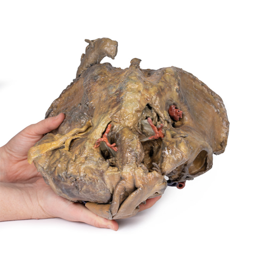
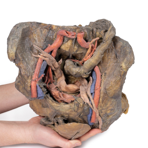
| 特長 | 本製品の説明は英語のみです。 Product information "Female pelvis deep dissection" This 3D model presents a deep dissection and isolation of the pelvis from surrounding regions, particularly demonstrating visceral and neurovascular structures relative to deep ligaments and osseous features. Within the false pelvis, the sigmoid colon descends on the left side of the specimen to the rectum, passing superficially across the pelvic brim and the passage of the common and external iliac artery and vein. Adjacent to the sigmoid colon are parts of the sigmoid arteries and superior rectal artery, resting superficial to the common iliac vessels and near the descending ureter. Anterior in the true pelvis is the collapsed urinary bladder, and between the bladder and rectum rests the uterus. The organ is partially covered in the broad ligament, with both the suspensory ligament of the ovary and round ligament have been separated and pulled away from the peritoneum on both sides to expose surrounding blood vessels. While the ovarian ligaments, round ligaments, uterine tubes and ovaries are trapped within the peritoneal fold of the broad ligament, the reduction in ovary size (common with advanced age) has rendered these indistinguishable in the model. Lateral to these organs, branches of the internal iliac artery can be identified – as well as a retained median sacral artery in the midline between the two common iliac arteries. On the left side only the uterine artery can be seen laterally. On the right side, the obturator, superior vesical, and uterine arteries can be observed. In addition, the origins of the inferior epigastric artery and vein can be seen arising from the external iliac vessels just prior to exiting the inferior abdominal cavity. On the right side of the preserved pelvis, the entire femur and thigh musculature has been removed to demonstrate the obturator membrane, the articular cartilage of the acetabulum and the transverse ligament of the acetabulum. Posteriorly the entire gluteal region has been dissected to expose the superior gluteal foramen and the origin of the superior gluteal artery. The sacrotuberous ligament has been removed to demonstrate the sacrospinous ligament, with some branches of the inferior rectal artery retained within the exposed ischoanal fossa. On the left side of the preserved pelvis the sciatic nerve has been maintained within the greater sciatic foramen, as has the sacrotuberous ligament. The ischioanal fossa mirrors that of the right side, where branches of the inferior rectal artery have been retained relative to the fibres of the pelvic diaphragm, and the integration of the external anal sphincter on the projecting external rectal surface. |
|---|---|
| 仕様 | エルラージマー社製 |
| 消耗品購入 | |
| 備考 | メーカー品番:MP1141 Erler-Zimmer GmbH & Co.KG の模型製品は、日本国内において株式会社京都科学の独占販売製品です。 |
| 医療機器クラス分類 | なし |
| 特定保守管理医療機器 | 該当なし |
| JANコード | なし |
| 更新年月日 | 2020年05月30日 |
