| 13232-000 | 64,350 (税別58,500)円 |
|---|
- 病理模型
- 頭部
- 血管
- 3Dプリント
EZ-233
| 13232-000 | 64,350 (税別58,500)円 |
|---|
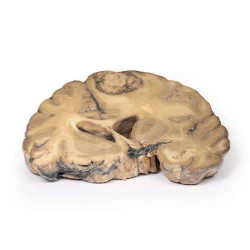
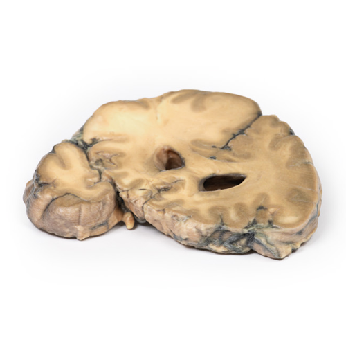
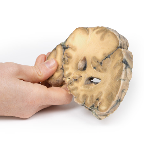
| 特長 | 本製品の説明は英語のみです。 Product information "Metastatic Adenocarcinoma in the Brain" Clinical History A 56-year old male underwent a total gastrectomy and splenectomy for gastric adenocarcinoma. Over a period of two months he developed a progressively unsteady gait, increasing weakness of his left hand and frontal headaches associated with nausea and vomiting. Imaging revealed a lesion in the right frontal lobe. He underwent a craniotomy with resection of the lesion, which was confirmed metastatic gastric adenocarcinoma. He experienced gradual increasing symptoms as well as jaundice, deteriorating consciousness and papilloedema from increased intracranial pressure. Repeat imaging revealed recurrence of the right frontal metastatic lesion as well as liver metastases. The patient died 9 months after his initial gastrectomy surgery. Pathology This brain specimen is cut in the coronal plane. A circumscribed, variegated, pink-grey tumour is evident in the right frontal lobe. The tumour is involving the grey and white matter. Compression of the right lateral ventricle by the lesion is apparent with shift of the midline structures also seen. Further Information Stomach cancer is one of the most common causes of cancer-related death worldwide. Risk factors include male gender, diet, smoking and chronic Helicobacter pylori infection. The most common sites for metastases of gastric adenocarcinoma are the liver, peritoneum, lungs and bones. Brain metastases are rare, occurring in <1% of cases. Isolated brain metastases are very uncommon with them being more commonly seen in disseminated disease and associated with a poor prognosis. Palliative treatment may include surgery, radiotherapy, steroid, chemotherapy or a combination thereof. |
|---|---|
| 仕様 | エルラージマー社製 |
| 消耗品購入 | |
| 備考 | メーカー品番:MP2007 Erler-Zimmer GmbH & Co.KG の模型製品は、日本国内において株式会社京都科学の独占販売製品です。 |
| 医療機器クラス分類 | なし |
| 特定保守管理医療機器 | 該当なし |
| JANコード | なし |
| 更新年月日 | 2020年06月16日 |
