| 13239-000 | 67,210 (税別61,100)円 |
|---|
- 病理模型
- 頭部
- 血管
- 3Dプリント
EZ-240
| 13239-000 | 67,210 (税別61,100)円 |
|---|
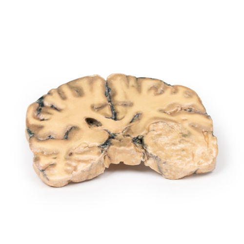
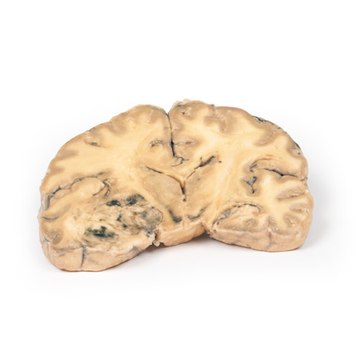
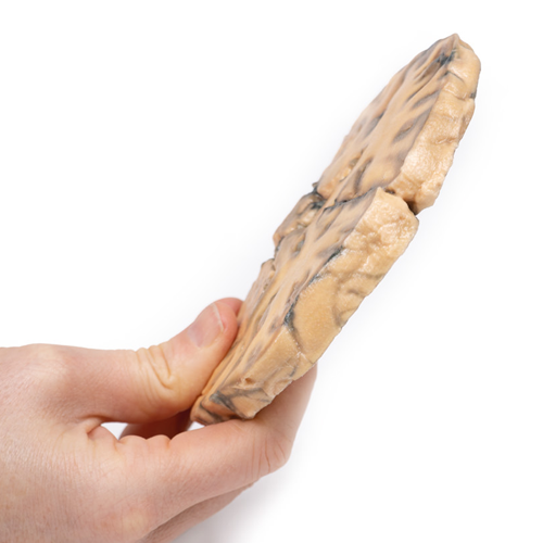
| 特長 | 本製品の説明は英語のみです。 Product information "Astrocytoma" Clinical History A 73-year-old female was admitted with new left-sided hemiplegia. On further questioning she revealed a 3-month history of headaches, nausea and deteriorating balance. CT brain revealed an inoperable brain tumour. She died 1 week after being admitted. Pathology This brain specimen is a coronal section. In the right temporal lobe, a poorly demarcated tumour is present. There is enlargement of the hemispheres and flattening of the gyral pattern. From the posterior aspect of the specimen subfalcine herniation* is appreciated and the tumour appear less well differentiated with haemorrhagic and necrotic foci. Histology of this tumour showed an astrocytoma, Grade III/IV. *In subfalcine (or cingulate) herniation, the most common type of brain herniation, the innermost part of the frontal lobe is pushed under part of the falx cerebri, between the two hemispheres of the brain. Further Information Gliomas are the second most common cancer of the central nervous system after meningiomas. The term “glioma” refers to tumours that are histologically similar to normal glial cells i.e. astrocytes, oligodendrocytes and ependymal cells. They arise from a progenitor cell that differentiates down one of the cell lines. Astrocytomas develop from the astrocyte lineage of glial cells. Tumours are staged according to histological differentiation and range from diffuse astrocytoma (Grade II/IV) to anaplastic astrocytoma (Grade III/IV) to glioblastoma (Grade IV). Histological features include the prominent eosinophilic cytoplasm in some astrocytic tumour cells (gemistocytes) as well as a fibrillary background. Astrocytomas occur most commonly between the fourth and sixth decades of life. Tumours usually occur in the cerebral hemispheres but may also occur in the cerebellum, brainstem or spinal cord. They most commonly present with seizures, headaches, nausea and focal neurological deficits depending on area involved. Without treatment Grade III median survival is 18 months. Treatment includes surgical resection, radiotherapy, chemotherapy or a combination thereof, depending on the clinical context. |
|---|---|
| 仕様 | エルラージマー社製 |
| 消耗品購入 | |
| 備考 | メーカー品番:MP2014 Erler-Zimmer GmbH & Co.KG の模型製品は、日本国内において株式会社京都科学の独占販売製品です。 |
| 医療機器クラス分類 | なし |
| 特定保守管理医療機器 | 該当なし |
| JANコード | なし |
| 更新年月日 | 2020年06月16日 |