| 13294-000 | 164,120 (税別149,200)円 |
|---|
- 病理模型
- 臓器
- 3Dプリント
EZ-295
| 13294-000 | 164,120 (税別149,200)円 |
|---|
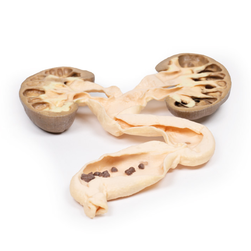
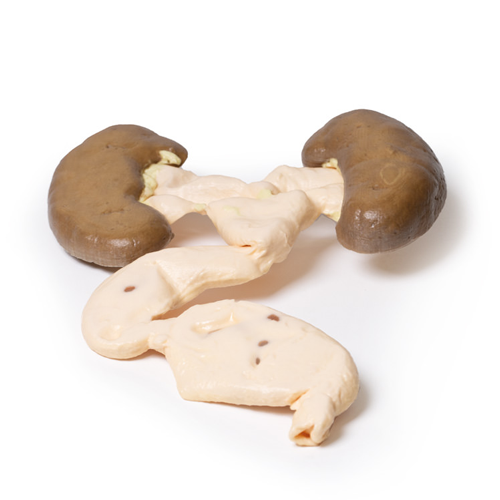
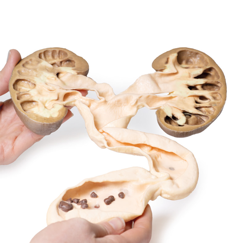
| 特長 | 本製品の説明は英語のみです。 Product information "Hydronephrosis Hydroureter" Clinical History A 49-year old male presents with a 6-week history of malaise, urinary frequency and haematuria for 6 weeks. Further questioning revealed intermittent left flank pain. Abdominal ultrasound showed severe hydronephrosis and hydroureter, secondary to multiple obstructing ureteric calculi at the uretero-vesical junction. He underwent a left nephrectomy and ureterectomy, and made a successful recovery. Pathology This is the patient’s left nephrectomy and ureterectomy specimen. The kidney has been bisected and the cut surface of both halves is displayed, mounted in continuity with the ureter, which has been opened. The kidney is grossly hydronephrotic, and there is considerable atrophic thinning and loss of renal parenchymal tissue. The ureter is extremely dilated and distally contains a number of small brown-black calculi with irregular sharp surface projections. These are calcium oxalate stones. This is an example of hydronephrosis and hydroureter due to calculi obstructing the lower end of the ureter. Further Information Hydronephrosis, or obstructive uropathy, is the dilation of the renal pelvis and calyces caused by an obstruction in the urine outflow. Obstruction can occur at any point in the urinary tract. Any lesion-, intrinsic (within the outflow system) or extrinsic (outwith the ureter)-, that impedes the flow of urine can lead to hydronephrosis. Common causes include: congenital anomalies, urinary calculi, urinary tract tumours, urinary tract inflammation, prostatic hypertrophy, and prostate tumours. Symptoms of the hydronephrosis relate to the pathology causing the obstruction (e.g. renal colic pain with calculi), the time period of the obstruction (acute or chronic), the site (unilateral or bilateral) and whether it is complete or partial. If the obstruction is not relieved it will ultimately cause pressure to build up proximal to the obstruction. This pressure is transmitted in a retrograde manner through the collecting ducts to the cortex causing progressive atrophy of the kidney with dilatation of the renal calyces and pelvis. The pressure also compresses the vasculature in the medulla leading to ischaemic medullary damage. Glomerular filtration persists in the affected kidney until late in the disease process when the filtration gradually diminishes or ceases. Obstruction triggers an interstitial inflammatory process leading to fibrosis. Ultrasound is the key diagnostic tool for diagnosis followed by CT or urogram. Most obstructing lesions require surgical intervention to relieve the blockage. Surgical interventions depend on each individual cause, but include nephrostomy or stenting for upper urinary tract obstruction and urinary catheter or suprapubic catheter insertions for lower urinary tract obstructions. |
|---|---|
| 仕様 | エルラージマー社製 |
| 消耗品購入 | |
| 備考 | メーカー品番:MP2093 Erler-Zimmer GmbH & Co.KG の模型製品は、日本国内において株式会社京都科学の独占販売製品です。 |
| 医療機器クラス分類 | なし |
| 特定保守管理医療機器 | 該当なし |
| JANコード | なし |
| 更新年月日 | 2020年06月01日 |
