| 13313-000 | 231,330 (税別210,300)円 |
|---|
- 病理模型
- 骨
- 下半身
- 3Dプリント
EZ-314
| 13313-000 | 231,330 (税別210,300)円 |
|---|
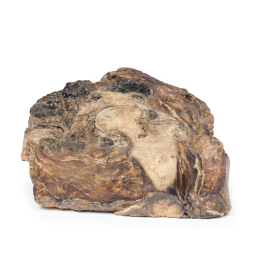
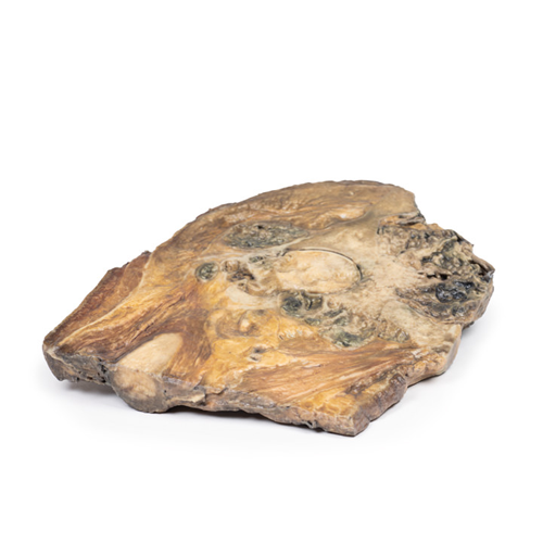
| 特長 | 本製品の説明は英語のみです。 Product information "Chondrosarcoma of femur and ilium" Clinical History A teenage boy presents with groin pain after horse-riding. Examination revealed a large, deep lump. Following biopsy and imaging, the diagnosis of chondrosarcoma was made and a radical surgical resection of his right leg was performed. Pathology The specimen consists of the upper end of the femur and its articulation with the pelvis. Within the neck and head of the femur and replacing most of the ilium there is a lobulated pale grey tumour with areas of cavitation, necrosis and haemorrhage. The tumour is extending out beyond bone into the surrounding soft tissues and appears encapsulated. The presence of infiltration, necrosis and haemorrhage are macroscopic features of malignancy. Further information Chondrosarcoma is a primary malignant bone tumour with cartilaginous differentiation. It is a rare cancer that accounts for about 20% of bone tumours. The only available treatment is excisional surgical resection since the current adjuvant treatments are ineffective. The pelvic location creates specific technical difficulties both for exeresis and reconstruction. The disease usually starts in the bones of the arms, legs or pelvis, but it can be found in any part of the body that contains cartilage. Sometimes chondrosarcoma grows de novo form an otherwise healthy bone; however, sometimes it may arise from a benign bone tumour (an enchondroma or osteochondroma). There are several subtypes of chondrosarcoma, named based on their microscopic and genetic characteristics. These include: conventional chondrosarcoma; Clear cell chondrosarcoma; Myxoid chondrosarcoma; Mesenchymal chondrosarcoma; Dedifferentiated chondrosarcoma. |
|---|---|
| 仕様 | エルラージマー社製 |
| 消耗品購入 | |
| 備考 | メーカー品番:MP2112 Erler-Zimmer GmbH & Co.KG の模型製品は、日本国内において株式会社京都科学の独占販売製品です。 |
| 医療機器クラス分類 | なし |
| 特定保守管理医療機器 | 該当なし |
| JANコード | なし |
| 更新年月日 | 2020年06月14日 |
