| 13317-000 | 96,910 (税別88,100)円 |
|---|
- 病理模型
- 骨格
- 3Dプリント
EZ-318
| 13317-000 | 96,910 (税別88,100)円 |
|---|
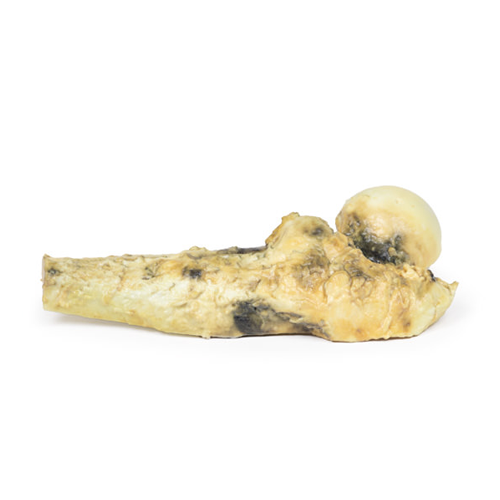
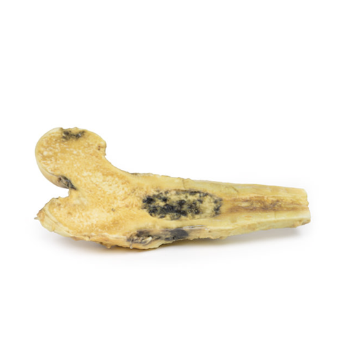
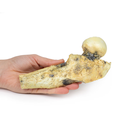
| 特長 | 本製品の説明は英語のみです。 Product information "Osteosarcoma of femur" Clinical History A 57-year old male attends complaining of recurrent pain in his right thigh. On examination there is no palpable abnormality in the thigh. An x-ray of the limb showed bony absorption associated with expansion and periosteal reaction at the proximal right femur. A CT of the limb showed a mass in the proximal right femur. A biopsy was taken of the lesion. Subsequently he had excision of the right upper femur followed by insertion of a prosthesis. Pathology The specimen comprises the head, neck and upper third of the shaft of the right femur, sawn longitudinally to display the cut surface. In the medullary cavity of the upper portion of the shaft is an ovoid tumour that is 6.5 cm in maximum diameter. The tumour is not encapsulated and has a haemorrhagic cut surface with pale hyaline and cystic areas. Histologically, this is a low grade chondrosarcoma. Further Information Chondrosarcomas are malignant bone tumours that produce cartilage. These are the third most common primary bone malignancy after myeloma and osteosarcoma. Conventional tumours are the most common subtype of chondrosarcoma, making up 90% of cases. Less frequently diagnosed subtypes include clear cell, dedifferentiated and mesenchymal chondrosarcomas. Some chondrosarcomas arise from pre-existing benign lesions, such as enchondroma or osteochondroma. Common mutations in chondrosarcomas are point mutations in the IDH1 and IDH2 genes as well as silencing of CDKN2A tumour suppressor gene. Chondrosarcomas that occur in multiple osteochondroma syndrome have mutations in the tumour suppressor EXT genes. Men are twice as likely to develop chondrosarcoma than women. The axial skeleton is more frequently affected than the appendicular skeleton. Around 20% affect the femur. These are largely slow growing tumours. They usually present with painful and gradually enlarging masses. At the time of diagnosis, most are low grade tumours that rarely metastasize. The lungs are the most common site for distant spread. Grade 1 tumours have an almost 90% 5-year survival rate, whereas with grade 3 chondrosarcoma the 5-year survival rate drops to 43%. CT scan is the optimal radiological investigation for diagnosis with MRI also frequently used. Biopsies may be taken to assist diagnosis. Treatment depends on the grade and the location of the tumour. Complete surgical resection is the standard treatment. Generally, chondrosarcomas do not respond to chemotherapy or radiotherapy given they very slow growing tumours. |
|---|---|
| 仕様 | エルラージマー社製 |
| 消耗品購入 | |
| 備考 | メーカー品番:MP2116 Erler-Zimmer GmbH & Co.KG の模型製品は、日本国内において株式会社京都科学の独占販売製品です。 |
| 医療機器クラス分類 | なし |
| 特定保守管理医療機器 | 該当なし |
| JANコード | なし |
| 更新年月日 | 2020年06月14日 |
