| 13319-000 | 140,690 (税別127,900)円 |
|---|
- 病理模型
- 骨格
- 3Dプリント
EZ-320
| 13319-000 | 140,690 (税別127,900)円 |
|---|
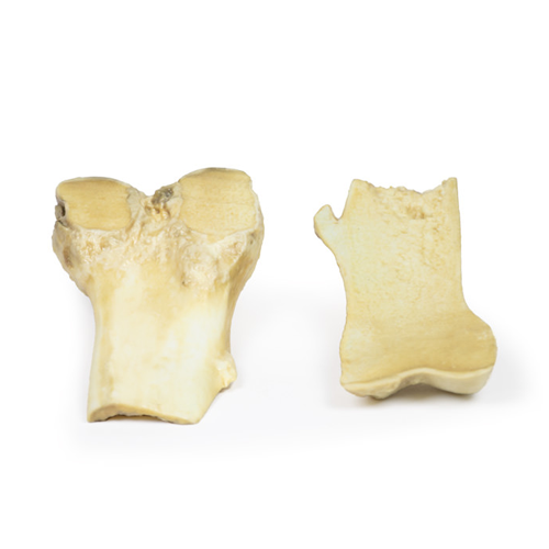
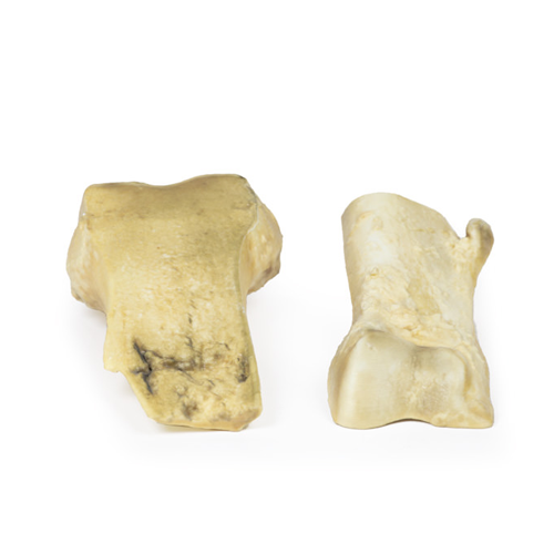
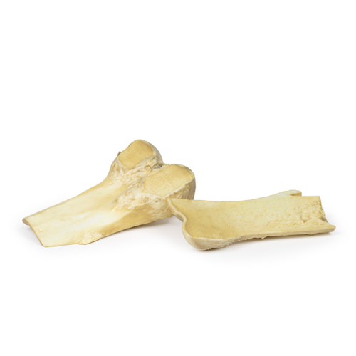
| 特長 | 骨軟骨腫(別名 外骨腫)は、骨腫瘍のなかで最も多い良性腫瘍です。 Product information "Osteochondroma" Clinical History A 61-year old male with prostate cancer attends pre-assessment clinic prior to a prostatectomy. Overall, he feels well with no major complaints. On review of symptoms, it is noted he has chronic pain in his right knee, which his GP called osteoarthritis. To exclude boney metastases of the prostate carcinoma, a knee x-ray is ordered, which shows a pedunculated lesion projecting from the medial aspect of the diaphysis of the right femur. His prostatectomy goes ahead but he subsequently dies from a postoperative pulmonary embolism. Pathology The specimen is the lower end of the patient’s right femur, which has been cut in the coronal plane and mounted to display the external surfaces. A pedunculated bony protuberance 2 cm in length projects from the medial aspect of the femoral shaft 7 cm above the medial condyle. The projection is composed of normal bone with a thin cap of hyaline cartilage at the tip. This is an example of an osteochondroma. Further Information An osteochondroma (or an exostosis) is a benign cartilaginous tumour. They are comprised of a cartilaginous capped bony protrusion from the external surface of the bone from which they arise. They are the most common benign bone tumours. Most osteochondromas occur spontaneously but they may also occur as part of multiple hereditary exostosis syndrome or post radiotherapy. They usually develop from or near the growth plate. They most commonly arise from the appendicular skeleton, especially in the lower limb around the knee or the upper limb at the proximal humerus. Men are more commonly affected than women. Symptoms vary on the site and size of the growth. Many osteochondroma remain asymptomatic. Osteochondroma lead to symptoms from the compression of surrounding neurovascular structures. They may also cause a pain from myositis or a fracture of the bony spur. They usually present in the second decade of life. They can be diagnosed with plain x-ray but MRI is the gold standard to ensure that there is no malignancy present within the growth. Hereditary exostoses are associated with mutations in the EXT1 and EXT2 genes. Reduced expression of these genes has also been seen in sporadic osteochondromas. Osteochondromas stop forming as fusion of the growth plate occurs. Treatment of excision is only if symptoms are severe. Malignant transformation to chondrosarcoma is rare in sporadic cases but more common in hereditary exostosis (5-20%). |
|---|---|
| 仕様 | エルラージマー社製 |
| 消耗品購入 | |
| 備考 | メーカー品番:MP2118 Erler-Zimmer GmbH & Co.KG の模型製品は、日本国内において株式会社京都科学の独占販売製品です。 |
| 医療機器クラス分類 | なし |
| 特定保守管理医療機器 | 該当なし |
| JANコード | なし |
| 更新年月日 | 2020年06月14日 |
