| Features |
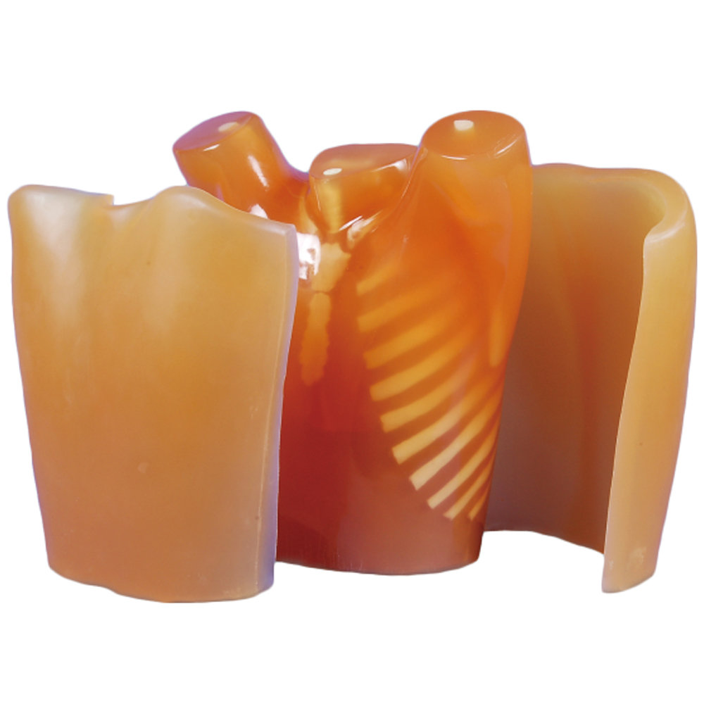
1. Radiation absorption and HU number approximate to human body, in an arms-abducted position suits the CT.
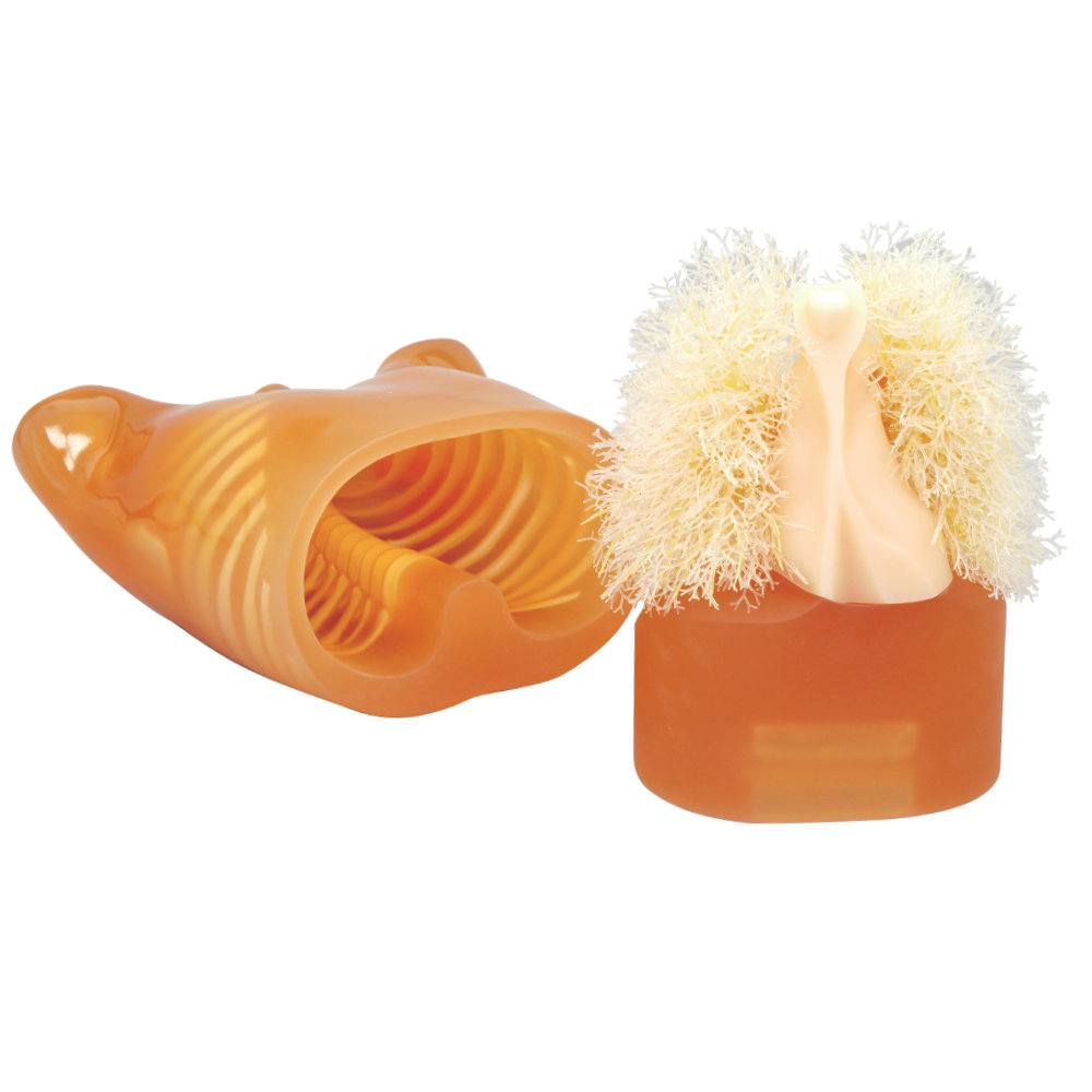
2.Simulated tumors and other targets can be attached at any points in the lung fields.
3. Wide variety of uses in interpretation training, anatomical education, evaluation and assessment of devices and other research. |
| Set includes |
(41337-100)
1 chest torso / 15 simulated tumors (15 variations 1 piece each) / 1 set of chest plates / 1 set of sample X-ray data (DVD) / manual
(41337-000)
1 chest torso / 15 simulated tumors (15 variations 1 piece each) / 1 set of sample X-ray data (DVD) / manual |
| Size (approx.) |
W43xH48 cm / W16.9xH18.9 in
chest girth: 94 cm / 37 in |
| Packing size (approx.) |
(41337-000)
W63 x D50 x H29cm / W24.8 x D19.7 x H11.4inch |
| Weight (approx.) |
18kg / 39.6lb |
| Packing weight (approx.) |
25kg / 55.1lb |
| Materials |
Soft tissue: urethane based resin (density: 1.06) / Synthetic bone: epoxy resin (density: 1.31) / *Phantom has no metal parts or liquid structure |
| Replacement parts |
41337-010 Chest Plates for PH-1 / 41363-020 Carrying case for PH-1 / 41337-070 N1 Simulated tumors for PH-1 |
| Related products |
PH-1C Pediatric Chest Phantom
PH-8 Lung Cancer Screening CT Phantom LSCT001
Breast plate for Chest Phantom N-1 LUNGMAN
Pneumonia module for Chest Phantom N-1 LUNGMAN
Components for Radioisotope
|
| Production & Development Supervision |
Kiyoshi Murata, Professor, Department of Radiology, Shiga Medical University / Norihisa Nitta, Director of Angiography Center, Kyoto Okamoto Memorial Hospital |
| Publication references |
Xie, X., Zhao, Y., Snijder, R. A., van Ooijen, P. M., de Jong, P. A., Oudkerk, M., ... Greuter, M. J. (2013). Sensitivity and accuracy of volumetry of pulmonary nodules on low-dose 16- and 64-row multi-detector CT: an anthropomorphic phantom study. European radiology, 23(1), 139-147. doi:10.1007/s00330-012-2570-7 / Gomi, T., Nakajima, M., Fujiwara, H., Umeda, T. (2011) Comparison of Chest Dual-energy Subtraction Digital Tomosynthesis Imaging and Dual-energy Subtraction Radiography to Detect Simulated Pulmonary Nodules with and without Calcifications. Academic Radiology, 18(2), 191-196. doi:10.1016/j.acra.2010.09.021 |
| Update |
July 26, 2020
|