Lung Sound Auscultation Trainer "LSAT" ver.2
Product supervision:
Chiharu Yoshii, M.D., Ph.D., Assistant Professor
Masamitsu Kido, M.D., Ph.D., Professor
Division of Respiratory Disease, University of Occupational and Environmental Health, Japan
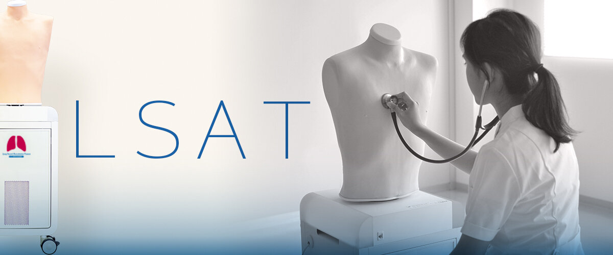
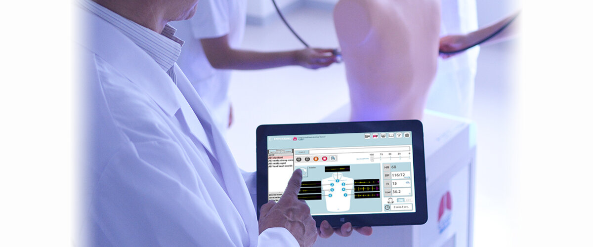
This unique and upgraded trainer can offer more efficient and effective respiratory auscultation training.
The physical status adjustable by touching control PC.
FEATURES
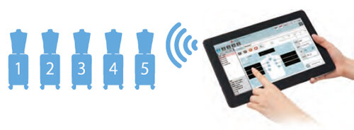
Wireless multi-unit operation
Up to five "LSAT" can be controlled b y one wireless control PC.
Each simulator can be individually programmed.
Cases can be switched at any time with a simple touch.
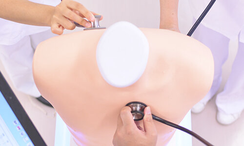
Simultaneous anterior and posterior auscultation
Two or more trainees can work together at the same by real stethoscopes.
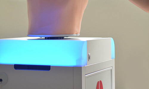
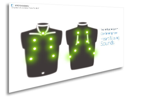
LED light panel to indicate inspiration and expiration
Attention to respiration rate while auscultating during examination
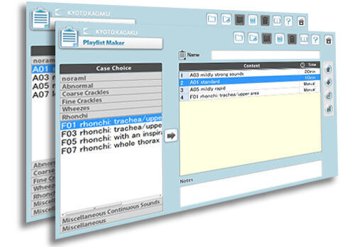
Create a custom playlist
With 35 different cases.
Incorporated "Playlist Maker" facilitates creating and conducting scenario based training sessions which feature change in finding over time.
Playlist Maker facilitates:
- sessions with accompany temporal change in physical findings
- standardizing training contents among different instructors.
- saving time of preparation
Error Indication System
The error indicator performs check-up of the system to keep LSAT in its best condition. Troubles and errors in speakers are warned on screen. History of system conditions is automatically recorded for reference.

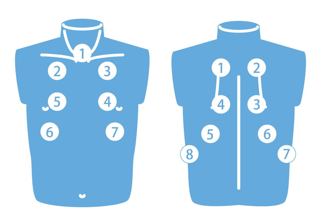
Basic Functions
 Left: Anterior / Right: Posterior
Anterior
1.trachea 2. upper right lung field 3. upper left lung field 4. middle left lung field 5. middle right lung field 6. lower right lung field 7. lower left lung field
Posterior
1. upper right lung field 2. middle right lung field 3. middle left lung field 4. lower left lung field 5. lower right lung field 6. right costophrenic angle 7. left costophrenic angle
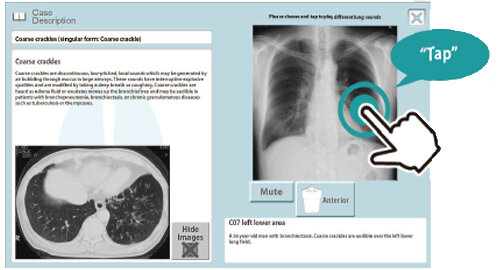
Case Information
Images of plain X-ray, CT and bronchoscopy are included.
Lung sound of each area can be played by tapping the plain X-ray image
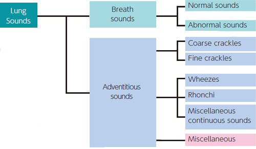
Classification of Lung Sounds
Based on acoustic analysis of recorded lungs sounds, each are classified according the American Thoracic Society standards. With this approach, lung sounds are categorized as continuous (wheezes, rhonchi, or stridor) or discontinuous (crackles). Crackles are further identified as fine or coarse.
CASES
Background heart sounds are available in 5 volume levels.
Normal
standard / mildly strong sounds / mildly rapid / loud heart sounds
Coarse crackles
right lower area / both lower area / right upper and middle area / left lower area / both upper area / whole area
Wheezes
upper area 600-700Hz / upper area 350-450Hz / upper area 200-1000Hz
Miscellaneous continuous sounds
stridors / squawks
Abnormal
weak: left lower area / weak: left whole area (adhesion) / weak: left whole area(pneumothorax) / absent: right middle and lower areas / weak: right whole area / absent: right whole area / weak: whole area / bronchial breathing
Fine crackles
both lower area / both middle lower area / whole area (IPF) / whole area (NSIP)
Rhonchi
trachea and upper area 150-250Hz / trachea and upper area 150-450Hz(polyphonic) / trachea and upper area 80-120Hz / whole area 80-200Hz
Miscellaneous
pleural friction rubs: right lower and middle area / pleural friction rubs: left lower area / Hamman's sign / Vocal fremitus