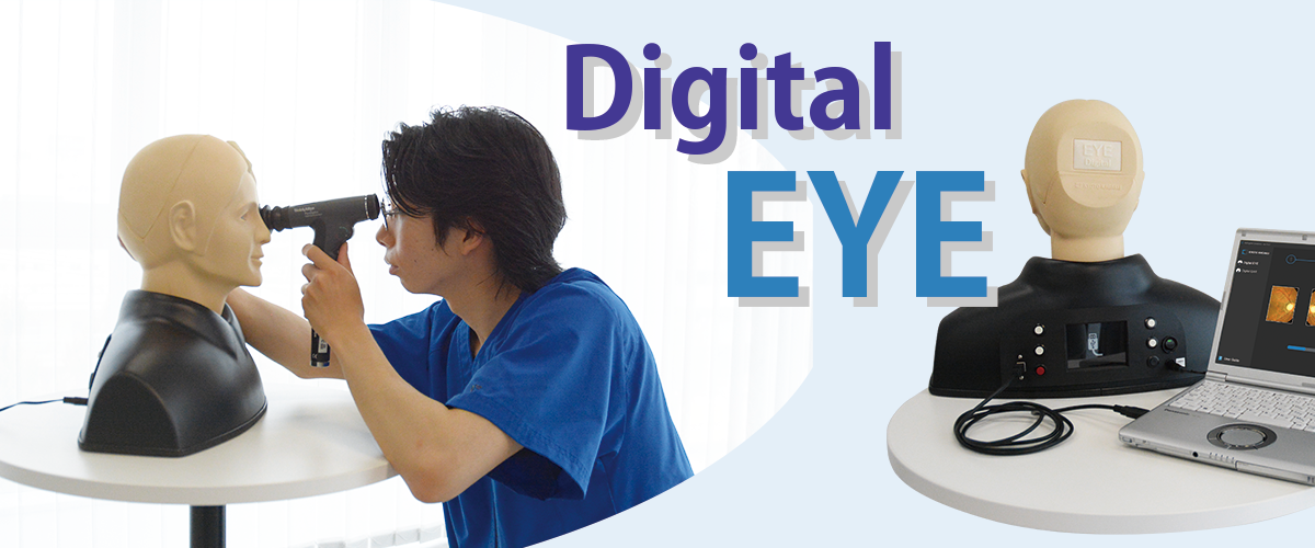
Digital EYE Examination Simulator
Product Supervision
Japan Society for Medical Education
with the cooperation of Michihiko Sone Professor - Nagoya university Graduate school of Medicine
Comparison

40 Pre-set Cases
-- Cases No.11–40: All-New Content --
- Normal fundus
- Hypertensive retinopathy
- Diabetic retinopathy
- Papilloedema (chronic phase)
- Papilloedema (acute phase)
- Glaucomatous optic atrophy
- Retinal vein occlusion (acute phase)
- Retinal vein occlusion (post retinal laser photocoagulation)
- Toxoplasmosis
- Age-related macular degeneration
- optic disc cupping 1
- optic disc cupping 2
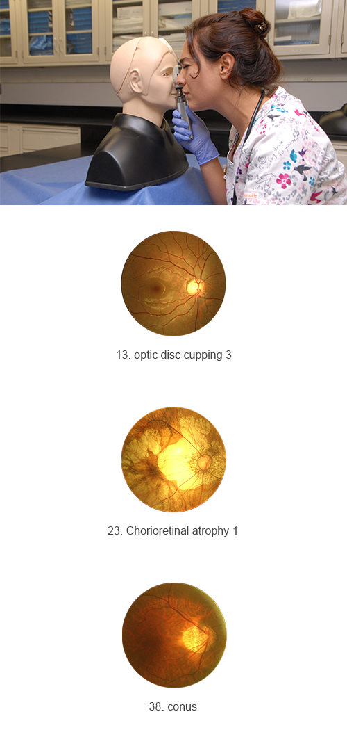
- optic disc cupping 3
- optic disc cupping, Tilted disc (Fuch's Coloboma)
- optic disc cupping, exudates
- Drusen 1
- Drusen 2
- Optic disc cupping with drusen 1
- Optic disc cupping with drusen 2
- Macular Drusen
- Optic disc hemorrhages
- Hemorrhages
- Chorioretinal atrophy 1
- Chorioretinal atrophy 2
- Vitreous clouding 1
- Vitreous clouding 2
- Vitreous clouding 3
- Vitreous clouding 4
- Retinal nerve fiber layer defect 1
- Retinal nerve fiber layer defect 2
- Pigmentation
- Hemorrhages, sheathing of vessel
- photocoagulation
- Megalopapilla
- Epiretinal membrane 1
- Epiretinal membrane 2
- Epiretinal membrane 3
- conus
- Macular Degeneration
- eye exudate
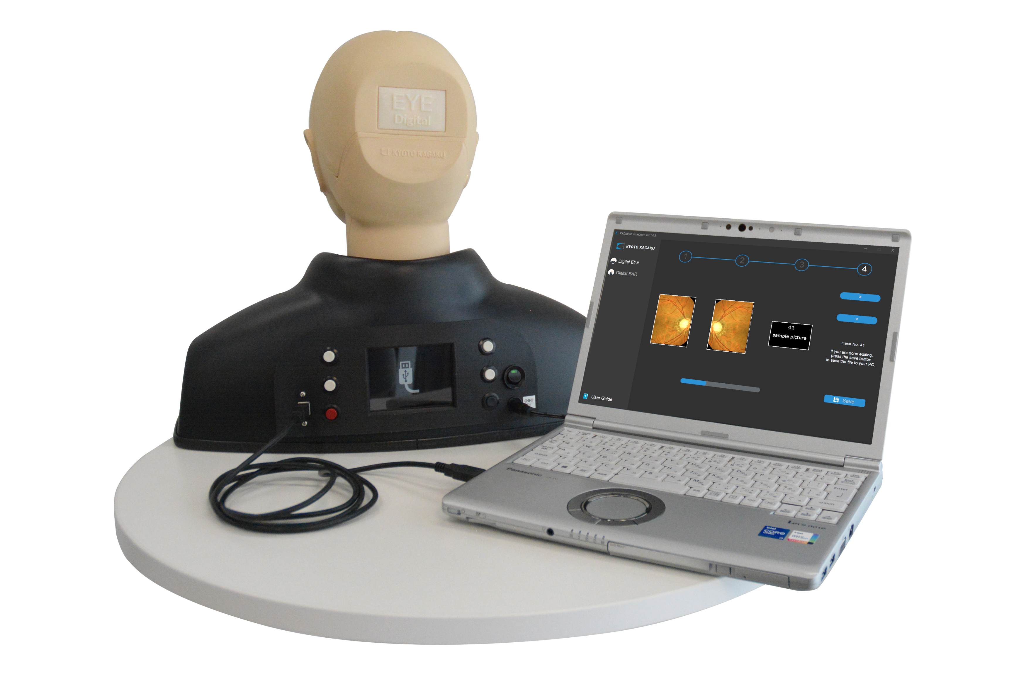
Case Install
Install Your Original Fundus Images
In addition to the 50 pre-installed images, you can import up to 50 of your own fundus images into the simulator via USB, using the case installer included in the set.
Easy to set up
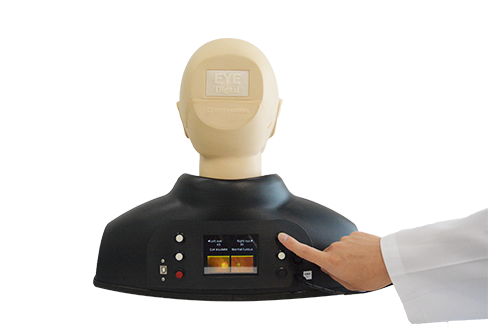
New design that is convenient for both instructors and learners.
- The case images can be switched with a single click of a button.
- The shown case can be monitored on the rear screen.
- Different cases can be displayed on the left and right eye.
Other Features (Shared with M82)
- Real clinical images
- The soft and supple material allows simulations of a real examination, in ways such as pulling up the eyelid
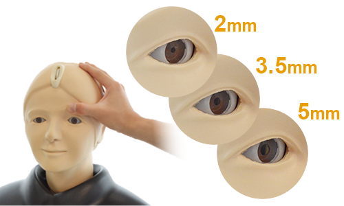
- 3 steps of contraction of the pupil (2, 3.5,5mm) (*MW61A is 2 steps; 3.5, 8mm)
- Training with a real ophthalmoscope