Fetus Ultrasound Examination Phantom SPACE FAN-ST Heart
Production & Development Supervision
Kiyoko Kabeyama RN, RM, PhD. Professor Midwifery & Women's Health, School of Human Health. Science, Graduate School of Medicine, Kyoto University
Haruto Egawa, PhD. National Hospital Organization Kyoto Medical Center. Department of Obstetrics, Head Physician or Medical Director
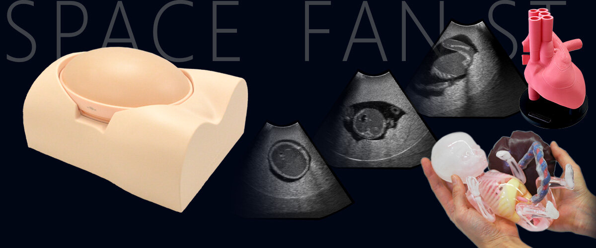
Now upgraded for fetal echocardiography training; Staple phantom for hands-on practice in second trimester screening
SKILLS
For fetal examination with ultrasound.
- Fetal size assessment: BPD, AD, AC and FL
- Measurement of amniotic fluid volume
- Determination of fetus presentation (cephalic or breech)
- Assessment of each body part
- Head: skull and brain
- Spine and limbs
- Cardiac chambers, blood vessels and lungs
- Assessment of umbilical cord and placenta position
- Determination of sex (This phantom represents a male fetus)
Designed with high anatomical fidelity to an actual fetus, allowing training with diverse measurement methods.
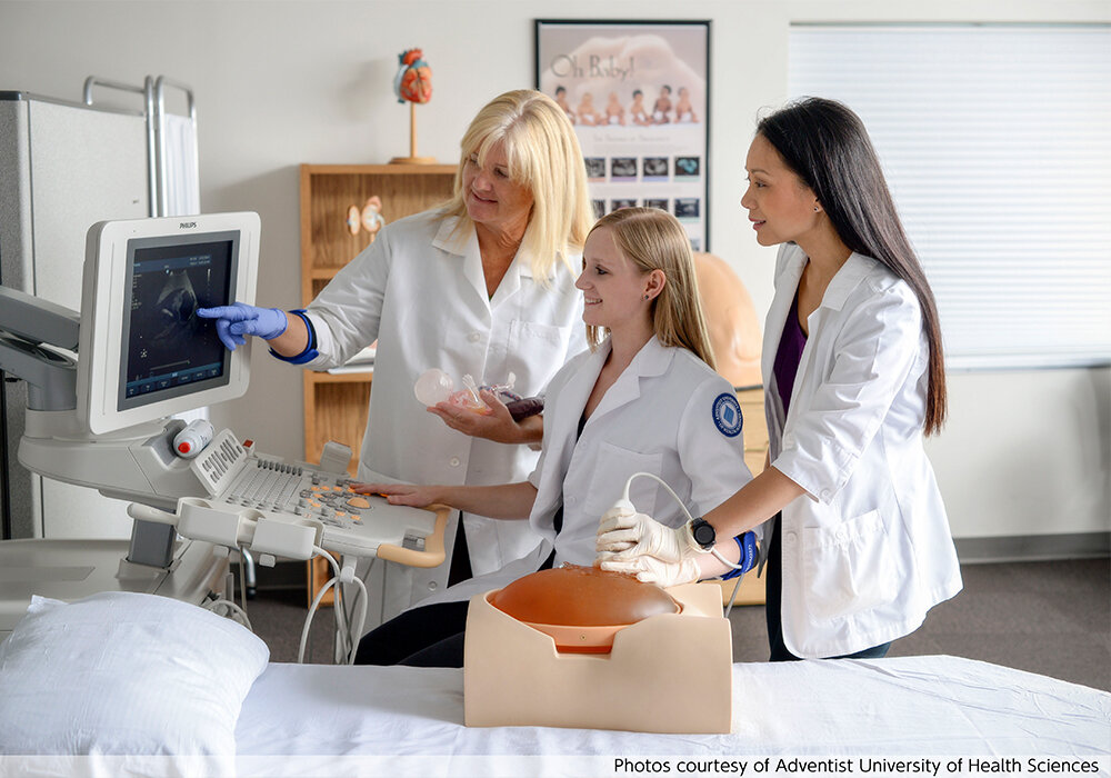
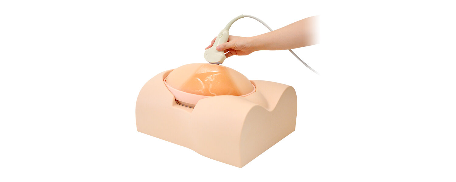 Usage image of the SPACE FAN
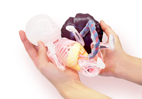 Fetus Demonstration Model (designed almost identically to the fetus embedded in the phantom)
Highly detailed fetus model
The fetus includes full skeletal structures and key organs
FETUS:
- skeletal structure
- brain with septum lucidum
- lateral ventricles and cerebellum
- heart with four chambers
- lungs
- spleen
- bladder
- stomach
- liver
- kidneys
- aorta
- umbilical vein
- umbilical artery
- external genitalia
UTERUS:
- amniotic fluid
- placenta
- umbilical cord
Fetus size: 26cm/10.24 in. (a 23-week fetus)
FEATURES
 Determination of fetus presentation / BPD / AC / FL / kidneys / heart / umbilical cord / umbilical vein
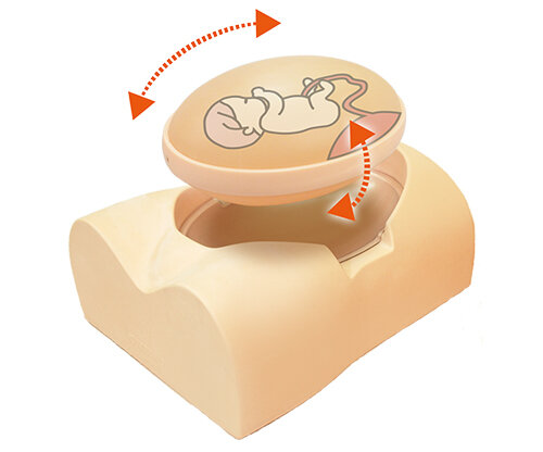
Anatomically Accurate Reproduction of the Intrauterine Environment
With actual ultrasound probes, trainees can practice screening examinations in a highly realistic manner.
1. Fetal Measurements
-
BPD (Biparietal Diameter): Measured at the level of the cavum septi pellucidi
-
AC (Abdominal Circumference): Measured at the level of the stomach, abdominal aorta, and umbilical vein
-
FL (Femur Length): Measured across the entire length of the femur
Estimation of fetal weight and assessment of growth
Support for other measurement methods
2. Amniotic Fluid Volume
Vertical depth measurement at the largest pocket (Maximum Vertical Pocket method)
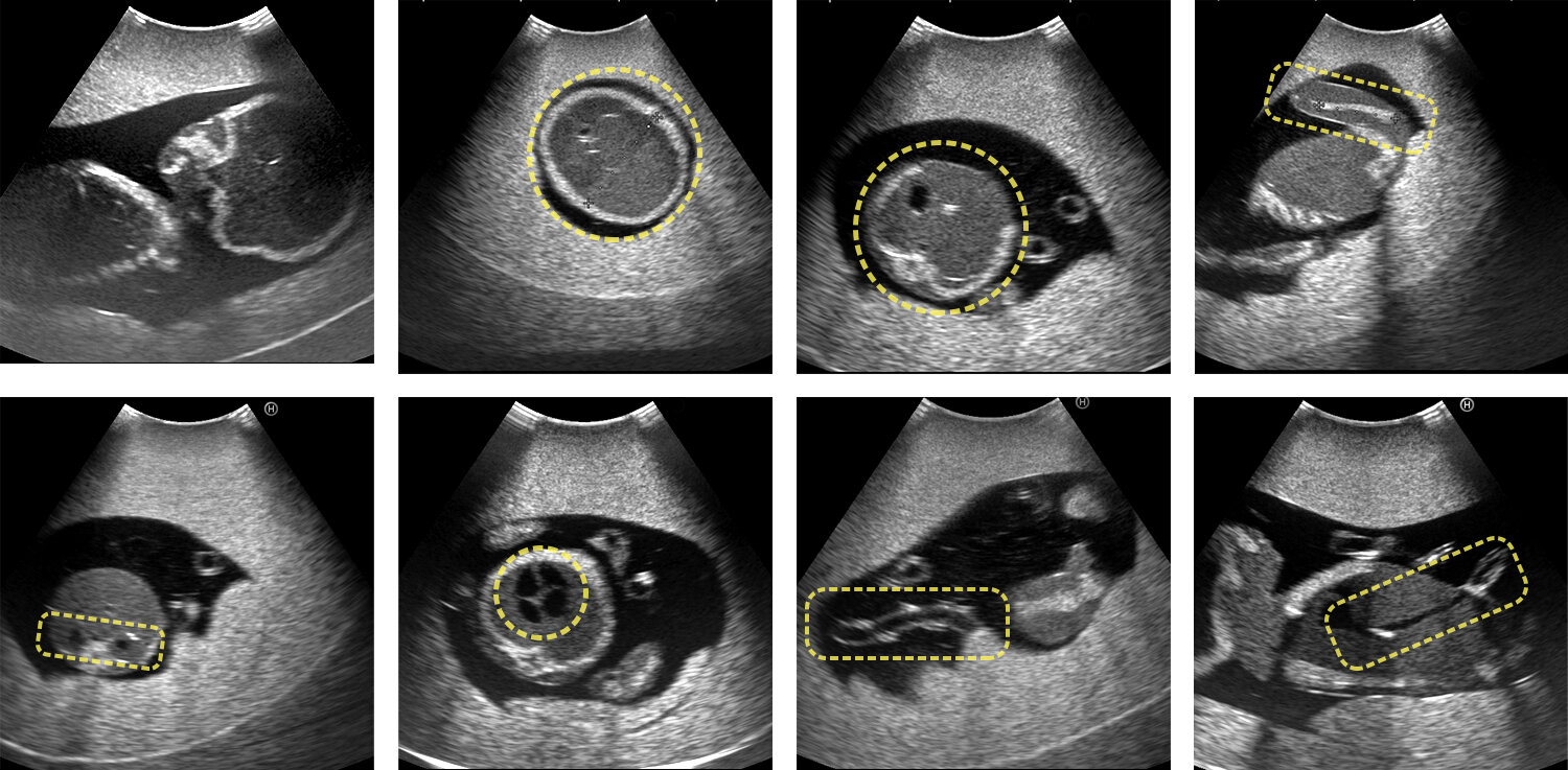
3. Diagnosis of Head, Thorax, Abdomen, and Spine
-
Skull morphology and brain assessment
-
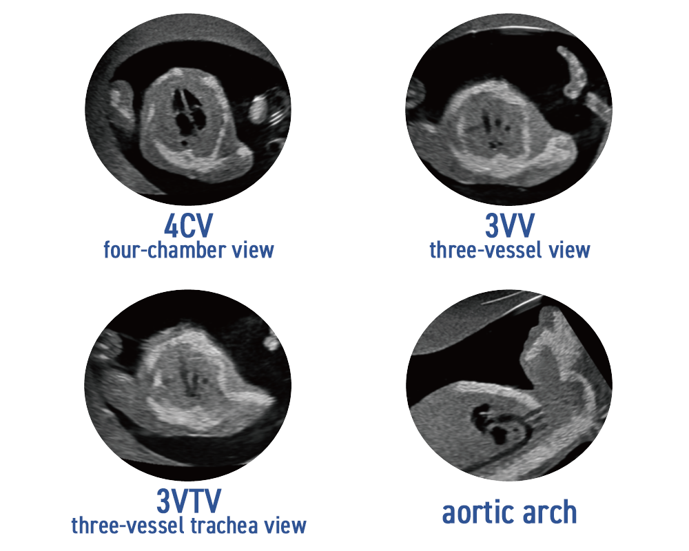
Heart four-chamber view, great vessels, inclination, and lungs
Note: Cardiac pulsation and blood flow dynamics are not reproduced. -
Abdominal organs and vasculature including stomach, kidneys, and bladder
-
Spine and limbs
4. Umbilical Cord and Placenta
Observation of the cord and its vessels, cord–placenta connection, and placental position.
5. Fetal Presentation
Determination of cephalic or breech presentation with interchangeable diagnostic section
6. Fetal Sex Determination
Reproduction of male genitalia for sex identification
Fetal Position & Orientation Made Easy!
The oval shape phantom abdomen can be set info four different positions.
Right: open hand, Left: fist
Detailed fetus heart
Detail echocardiography training
US-7B SPACE FAN-ST HEART for Fetal Echocardiography features detailed cardiac anatomy, ideal for echocardiography training.
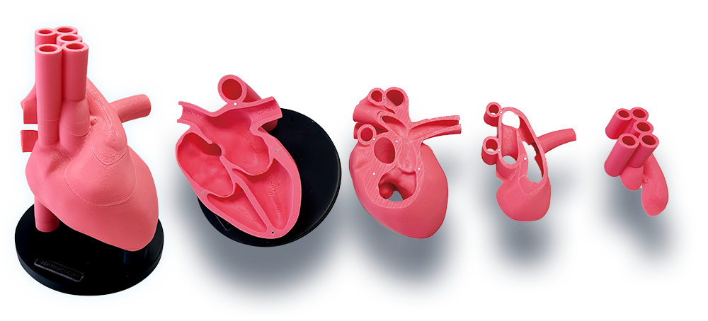
Fetus Heart Model
Three times enlarged model
Including magnified fetal heart model comes apart into 5 pieces according to the basic planes in fetal echocardiography. Can be taken apart to explore the internal anatomy.
Supporting Materials
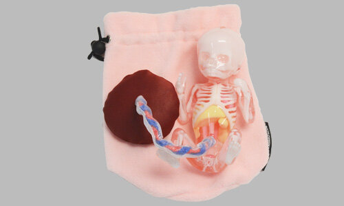
Fetus Demonstration Model
Life-size fetus model for demonstration and visual understanding
-
Enhances learning effectiveness when used in explanations for beginners
-
Helps trainees visualize fetal orientation during ultrasound imaging
-
Skull partially opened to allow observation of the transparent septum
-
Placenta with detachable umbilical cord included
-
Comes with a fabric pouch for convenient storage
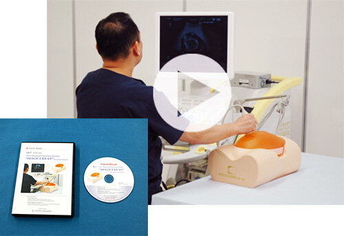 DVD image
DVD Included
SThis DVD presents the complete workflow of fetal ultrasound screening with SPACE FAN-ST. Supervised by Dr. Egawa, the DVD features clear and thorough explanations.
Contents:
-
Observation of the fetal head
-
Observation of the skeleton
-
Observation of the thoracic and abdominal regions
-
Observation of other structures
-
Observation of fetal appendages
-
Measurement methods
-
Breech presentation